Early Diagnosis of Cancer Based on Protein Microarrays
Keywords: protein chip, surface chemistry, biomarkers, cancer diagnosis, biosensor, silane, polymer, XPS, ATR-FTIR, ToF-SIMS, Contact Angle, Fluorescence scanning;
In collaboration with Departement bioMarqueurs of bioMérieux company , and two French Central Hospitals (CHU Montpellier, CHU Saint-Etienne)
Cancer is the first leading cause of death in the world, but the chance of cure will substantially increase if it can be precisely detected at an early stage. It has been observed that the sera of cancer patients contain autoantibodies against tumor-associated antigens (TAAs), which can be detected before clinical symptoms occur. However, the low frequency and heterogeneity of TAAs in patients’ sera bring challenges to traditional detection methods (e.g. enzyme linked immunosorbent assay, ELISA), especially some autoantibody lacking specificity. Protein chip technology provides a useful approach for the simultaneous serodetection of various antibodies with high throughput and tiny volume requirements. The crucial parameter of protein chip is design of surface chemistry to well immobilize proteins and retain their biological activities. Besides, the concentration of probe proteins, spotting buffer (pH, additives), capping and incubation conditions should be also taken into account for optimisation.
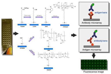
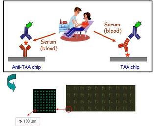
We fabricated 3D micro-structure glass slides (Fig.1) with photolithography technique for the solid support of protein chip. The microstructured slides not only retains surface properties of the original glass substrate, but also do not increase the fluorescence background level, which allows to incubate with each target sample (antigen, antibody, serum) per well and each surface chemistry per well. The surface-coating was performed with silanes (amino, epoxy, and carboxylic), or polymers layers: Jeff amine, chitosan, carboxymethyl dextran (CMD, synthesized in our lab), maleic anhydride-alt-methyl vinyl ethercopolymer (MAMVE) for physicochemical adsorption with proteins. Moreover, we have optimized the parameters, such as concentration of probe proteins, spotting buffer (with various pH and additives), capping and incubation conditions, in order to implement customized protein microarray. At last, the low cost, high-throughput and high sensitive technique was employed to evaluate biomarkers involved breast and colorectal cancer in serum of patient for clinical profiles (Fig.2).
Label-free Microcantilever Sensors for Point-of-Care Cancer Diagnosis
Keywords: Microcantilever, surface stress, surface chemistry, phase-shifting interferometric microscopy (PSIM), cancer biomarkers
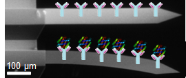
Molecules adsorbed, and biochemical reaction, on a functionalized microcantilever cause vibrational frequency changes and static deflections of the microcantilever, and can be sensitively measured by detecting changes like surface deflection (Fig. 3). Basically, a cantilever sensor substrate has two different sides: one side (typically the upper surface) is coated with receptor molecules (like antibodies, ssDNA etc) for specific adsorption to measure the surface-stress-induced deflection of a cantilever. The other surface of the cantilever (typically the lower surface) may be left uncoated, or be coated with a passivation layer which is inert or does not exhibit significant affinity to the analyte in order to limit non-specific adsorption. The natural silicon dioxide layers on silicon and coated gold surfaces are commonly used as the two sides of cantilever sensor substrates.
Molecules adsorbed, and biochemical reaction, on a functionalized microcantilever cause vibrational frequency changes and static deflections of the microcantilever, and can be sensitively measured by detecting changes like surface deflection (Fig. 3). Basically, a cantilever sensor substrate has two different sides: one side (typically the upper surface) is coated with receptor molecules (like antibodies, ssDNA etc) for specific adsorption to measure the surface-stress-induced deflection of a cantilever. The other surface of the cantilever (typically the lower surface) may be left uncoated, or be coated with a passivation layer which is inert or does not exhibit significant affinity to the analyte in order to limit non-specific adsorption. The natural silicon dioxide layers on silicon and coated gold surfaces are commonly used as the two sides of cantilever sensor substrates.
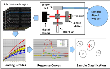
We try to detect cancer biomarkers using micro cantilever (MC) sensors based on our home-made readout system of phase-shifting interferometric microscopy (PSIM), which can sensitively measure displacement of surface stress in nm range or below. Compared to existing cantilever-sensor readout methods, PSIM does not depend on alignment and allows for the monitoring of the entire displacement profiles of all cantilevers within the field of view, but using a single light source. Moreover, a sample cell which can hold multiple cantilever-array chips has been developed, allowing rapid and reproducible sensor-chip replacement, individual or common addressing of all chips in the sample cell and very small sample requirement (Fig. 4). This can be potentially developed as a handheld biosensor for point-of-care diagnosis due to the possible miniaturised optical system.
Featured References
1. Characterization of three amino-functionalized surfaces and evaluation of antibody immobilization for the multiplex detection of tumor markers involved in colorectal cancer.
Yang Z, Chevolot Y, Gehin T, Dugas V, Xanthopoulos N, Laporte V, Delair T, Attaman Y, Choquet-Kastylevsky G, Souteyrand E,
Laurenceau E.
Langmuir 2013 29 (5): 1498–1509 PDF
2. Improvement of protein immobilization for the elaboration of tumor- associated antigen microarrays:Application to the sensitive and specific
detection of tumor markers from breast cancer sera.
Yang Z, Chevolot Y, Gehin T, Solassol J, Mange A, Souteyrand E, Laurenceau E.
Biosens Bioelectron 2013; (40)1: 385–392.PDF
3. Cancer Biomarkers Detection Using 3D Microstructured Protein Chip: Implementation of Customized Multiplex Immunoassay.
Yang Z, Chevolot Y, Attaman Y, Choquet-Kastylevsky G, Souteyrand E, Laurenceau E.
Sens Actuators B: Chem 2012,175: 22-28. PDF


